by Markus Gschwind
The patient was a 51-year-old car mechanic, without any previous illness apart from untreated arterial hypertension. One morning, on waking very early to go to work, he felt a strange dizziness. During breakfast he fell asleep twice while talking to his wife, and she observed his right eyelid was dropped so that the eye was not as open as usual. Despite her worries, he nevertheless took the car to work. Just before getting into his car, however, he fell forward onto the ground. While driving he hit the left side of the road several times, but never stopped, it was only when he lost his left hubcap that he stopped to retrieve it. Again, when trying to pick it up, he fell to the ground. At work his colleagues stared at him and asked “Have you seen your nose”? They noticed that the left corner of his mouth was drooping, so stopped him working and took him to the emergency department against his will.
The admitting physician found a bradycardia at 46 bpm, miosis with ptosis on the right and a left sided central facial paralysis. There was a discrete overshoot on the left side during cerebellar testing, the patient rotated 40° to the left in a stepping test and deviated to the left side on heel-toe walking.

Comment by the author
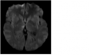
MRI scanning showed a new right thalamic ischemic stroke in the territory of the right tuberothalamic artery (VL and MD nuclei); (NIHSS =2: facial paralysis and ataxia).
This case serves as a nice anecdote illustrating the kaleidoscope of possible symptoms and signs possible in the context of a right thalamic stroke. While several of them have been reported separately, cases that include excessive sleepiness, ipsilateral Horner’s syndrome, contralateral facial paralysis, contralateral hemi-ataxia, contralateral hemi-neglect, and anosognosia as our patient did are rare.
Markus Gschwind is Medical Assistant at the Department of Clinical Neurosciences chaired by Professor Richard Frackowiak, CHUV Lausanne, Switzerland
Comment by Alessandro Padovani and Alessandro Pezzini
This male patient presented at the age of 51 with fluctuating consciousness, ipsilateral Horner’s syndrome with miosis and ptosis, contralateral facial paresis, hemispatial neglect and hemi-ataxia due to a right thalamic paramedian infarct as documented by MRI. Onset was sudden and the patient suffered arterial hypertension, thus making the suspect of ischemic stroke the most likely as thalamic infarcts both in isolation and in combination with infarcts involving other structures represent a frequent clinical entity (1); other less frequent possibilities include deep venous system thrombosis, Epstein-Barr virus encephalitis, cytomegalovirus encephalitis, diencephalic gliomas, metabolic disorders such as thalamic extrapontine lesions in central pontine myelinolysis or Wernicke encephalopathy, though these are generally associated to bilateral involvement.
This case follows a long series of case reports showing the myriad of signs and symptoms variably associated with focal thalamic lesions (2). In fact, the thalamus is a diencephalic structure whose numerous nuclei represent a complex gateway for input and output to and from the cerebral cortex. It receives, modifies, and relays information from major afferent, somatomotor, reticular formation, and limbic system pathways.
The author claims for a stroke in the territory of the right tuberothalamic artery, confirming previous isolated case reports similar to this. However, similar cases have been also associated to strokes in the territory of the paramedian arteries, thus casting some doubts on the involved vessel in the absence of other instrumental findings (MRA or cerebral angiography) and other neuropsychological details.
Four different patterns of clinical syndromes have been described (3, 4), reflecting the major thalamic vascular territories (tuberothalamic, paramedian, inferolateral, and posterior choroidal). However, there is plenty of evidence that neurological syndromes can be rather heterogeneous as consequence of the high variability of vascular supply of the anterior lateral and inferior medial thalamic nuclei (5).Variations of vascular supply of the thalamus and of extent of collateralisation are frequent, thus making the attribution of individual vascular occlusion patterns to a constellation of clinical deficiency symptoms and infarcts demonstrated by neuroimaging rather difficult as this original and interesting case seems to confirm.
1. Pezzini A, Del Zotto E, Archetti S, Albertini A, Gasparotti R, Magoni M, Vignolo LA, Padovani A.. Thalamic infarcts in young adults: relationship between clinical-topographic features and pathogenesis. Eur Neurol. 2002;47(1):30-6
2. Dejerine J, Roussy G. Le syndrome thalamique. Rev Neurol (Paris). 1906;14:521-532
3. Graff-Radford NR, Damasio H, Yamada T, Eslinger PJ, Damasio AR. Nonhaemorrhagic thalamic infarction: clinical, neuropsychological and electrophysiological findings in four anatomical groups defined by computerized tomography. Brain. 1985;108:485-516
4. Bogousslavsky J, Caplan LR. Vertebrobasilar occlusive disease: review of selected aspects—thalamic infarcts. Cerebrovasc Dis. 1993;3:193-205.
5. Schmahmann JD. Vascular Syndromes of the Thalamus. Stroke 2003, 34:2264-2278:
Alessandro Padovani is Full Professor of Neurology at the Department of Clinical and Experimental Sciences, University of Brescia, Italy
Alessandro Pezzini is Assistant Professor of Neurology at the Department of Clinical and Experimental Sciences, University of Brescia, Italy
Comment by Stefano Cappa
This is an intriguing case report. The clinical features of thalamic stroke according to the involved vascular territory have been well delineated (Schmahmann, 2003). In particular, the clinical picture of tuberothalamic territory involvement has been described in detail on the basis of a large patient series (Bogousslavsky et al., 1986). While hemispatial neglect is frequently observed after right sided involvement of this territory, the most striking feature of this case is the patient’s behavior, suggesting a defective awareness, not only of the right hemispace, as indicated by his driving, but also of several other worrisome events, such as the eyelid drop observed by the wife, and the two falls. This may lead to some considerations about the usage of the term “anosognosia”. This clearly refers to the defective awareness of a deficit, as indicated by overt verbal denial. An alternative possibility is that the patient may be aware of the deficit, but not showing the appropriate emotional reaction. Actually, what has been labeled as “emotional unconcern” was suggested to be one of the crucial features of tuberothalamic infarction (Lisovoski et al., 1993). The association with bradycardia is also worth mentioning. Life-threatening bradycardia has been recently reported in a patient with a bilateral paramedian thalamic and midbrain infarction (Perruzzotti-Jametti et al., 2011). A possible speculation is that a disconnection mechanism may be involved in both the behavioral disorder and the bradycardia, resulting in a separation of sensory information from the appropriate visceral emotional responses. As correctly underlined by the title of the case report, the thick packaging of nuclei and fibre tracts in the small volume of the thalamus can give rise to the most kaleidoscopic combination if signs and symptoms observed in clinical neurology.
Bogousslavsky J, Regli F, Assal G. The syndrome of unilateral tuberothalamic artery territory infarction. Stroke. 1986 May-Jun;17(3):434-41.
Lisovoski F, Koskas P, Dubard T, Dessarts I, Dehen H, Cambier J. Left tuberothalamic artery territory infarction: neuropsychological and MRI features. Eur Neurol. 1993;33(2):181-4.
Peruzzotti-Jametti L, Bacigaluppi M, Giacalone G, Strambo D, Comi G, Sessa M. Life-threatening bradycardia after bilateral paramedian thalamic and midbrain infarction. J Neurol. 2011 Oct;258(10):1895-7.
Schmahmann JD. Vascular syndromes of the thalamus. Stroke. 2003 Sep;34(9):2264-78.
Stefano Cappa is Professor of Cognitive Neuroscience at the San Raffaele University, Milan, Italy and past-chairman of the EFNS Scientist Panel Cognitive Neurology


2 comments
I know someone who had a right thalamic infarct and feels they have deficits with grasping with the left hand. They often drop things or misjudge where the object is in space or do not grip the object with enough strength to hold onto it. There have also been very frightening panic attack like seizures and deja vu, jamais vu that seem better controlled on Keppra. In fact this is how the infarct was detected on MRI when scanning for signs of epilepsy. Could the infarct and the seizures be connected? It seems to me very possible, but the data seems to be very limited.
Seizures are considered to be uncommon after thalamic hemorrhage, and extremely unusual after subcortical infarction (se, however, Giroud et al., Epilepsia, 35, 959,1994). In the case in question, however, one could speculate that the manifestations could reflect a sensory-limbic hyperconnection…