by Mélisande Rouger
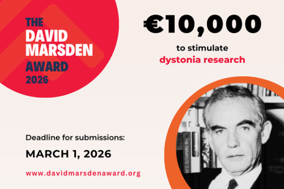
The popular Basal Ganglia Club reconvened again at EAN 2023 in Budapest, featuring both the MDS-ES C. David Marsden Award Lecture, delivered by Prof. Claudia Trenkwalder from the Department of Neurology, Paracelsus Elena Klinik, Kassel, Germany, and the Dystonia Europe David Marsden Award Lecture, given by Michael Zech from Munich, Germany.
The European Basal Ganglia Club is an established fixture of EAN Annual Congress, providing a reliable source of updates on therapy and management strategies of Parkinson’s Disease.
Prof. Trenkwalder, who is Past President of the International Parkinson and Movement Disorders Society, gave a comprehensive overview of the challenges in detecting and assessing isolated REM sleep behaviour disorder (iRBD).
“IRBD is currently the most specific prodromal feature of α-synuclein pathology, which may manifest even a decade before motor symptoms of Parkinson’s disease, dementia with Lewy Bodies (DLB) or multiple system atrophy (MSA) start,” she said.
The condition first appeared in the literature in 1986, as researchers from the United States described a new kind of parasomnia, which they called REM sleep behaviour disorder because patients presented with violent movements in their REM sleep.
“Those researchers put together a cohort of patients and followed them for many years,” she said. “After 16 years, more than 80% of these patients had developed a neurodegenerative disease, either Parkinson’s disease, DLB or MSA.”
The first description was later published in Sleep Medicine, showing that in the long term, this syndrome may convert to neurodegeneration.
Unique phenomenology
Trenkwalder then proceeded to describe the phenomenology of REM-sleep behaviour disorder, which is characterised by dream enactment, and reviewed the current options to assess and scale the disorder. “In normal REM sleep, we observe a complete relaxation of all muscles, no movement, no vocalisation and maybe some twitches in leg muscles,” she said.
By contrast in iRBD, an increased muscle tone can be observed, especially in the chin, she added. “Several methods for scaling this increased muscle tone have been developed by different groups, including the American Academy of Sleep Medicine, and teams from Montreal, Barcelona, and Innsbruck.”
The most specific sign of REM without atonia is measured in the chin region any time of this muscle tone in the REM sleep, she explained.
“REM without atonia is the only objective biomarker we have to measure RBD,” she said. RBD diagnostic clinical criteria as defined by the International Classification of Sleep Disorders from 2014 include repeated episodes of sleep related vocalisation and/ or complex motor behaviours.
“These behaviours that occur during REM sleep or, based on clinical history of dream enactment, are presumed to occur during REM sleep, are documented by polysomnography,” she said. “Polysomnographic recording demonstrates REM sleep without atonia. The disturbance is not better explained by another sleep disorder, mental disorder, medication, or substance use.”
RBD episodes can be mild with jerky movements of the upper extremities, up to complex movements of the whole body with a risk of injury. As for RBD pathways, REM-on neurons are glutematergic and excite GABA and glycinergic neurons with inhibition of spinal motor neurons causing REM atonia.
“In RBD, we observe a lack of inhibition of spinal motor neurons and thalamocortical neurons,” she said. To assess RBD, screening questionnaires have been developed form the Marburg and Innsbruck groups, but these questionnaires are not specific.
“It’s very difficult to detect RBD because patients are asleep,” she said. “Diagnosis cannot be reliably made by screening questionnaires; only probable RBD.” Assessment and diagnosis of iRBD with history also fail, as patients rarely remember the nocturnal events and bed partners’ descriptions are crucial, she added.
A more recent project with improved automatic identification of iRBD with a 3D time-of-flight camera, which shows promise was also discussed. ‘This test might be implemented in clinical routine as a supportive screening tool for identifying people with RBD,’ she said.
The Marburg group has followed isolated RBD patients with a reduced sympathetic cardiac innervation progress in the z-score of fluoro-deoxy-glucose-positron-emission-tomography (FDG-PET) to detect typical Parkinson’s patterns in the PET.
‘They found that only those patients with iRBD who had already reduced xxx pet at the baseline of diagnosis develop a typical Parkinson’s pattern on FDG PET after 4 to 5 years,’ she said.
The Dystonia Europe David Marsden Award Lecture was then given by Michael Zech from Munich, Germany.
The session was rouned off with four video case presentations, chosen through an application and selection process in collaboration with the International Parkinson and Movement Disorder Society – European Section, from Dr. Alvaro Bonelli from Madrid, Spain; Dr. Andona Milovanovic from Belgrade, Serbia; Dr. Sandy Cartella Reggio from Calabria, Italy; and Dr. Andres Fernandes from Porto, Portugal.


