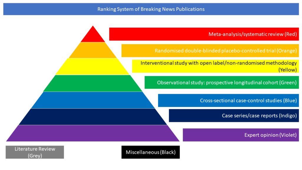
Cross-sectional case-control studies (Blue)
Read on for our pick of Covid-related cross-sectional case control studies from the scientific press for June 2022:
- Cerebral Microbleeds Assessment and Quantification in COVID-19 Patients With Neurological Manifestations
- Widespread white matter oedema in subacute COVID-19 patients with neurological symptoms
- Global Impact of the COVID-19 Pandemic on Cerebral Venous Thrombosis and Mortality
Cerebral Microbleeds Assessment and Quantification in COVID-19 Patients With Neurological Manifestations
It is increasingly acknowledged that Coronavirus Disease 2019 (COVID-19) can have neurological manifestations, and cerebral microbleeds (CMBs) have been observed in this setting. The aim of this study was to characterize CMBs patterns on susceptibility-weighted imaging (SWI) in hospitalized patients with COVID-19 with neurological manifestations. CMBs volume was quantified and correlated with clinical and laboratory parameters. The study included patients who were hospitalized due to COVID-19, exhibited neurological manifestations, and underwent a brain MRI between March and May 2020. Neurological, clinical, and biochemical variables were reported. The MRI was acquired using a 3T scanner, with a standardized protocol including SWI. Patients were divided based on radiological evidence of CMBs or their absence. The CMBs burden was also assessed with a semi-automatic SWI processing procedure specifically developed for the purpose of this study. Odds ratios (OR) for CMBs were calculated using age, sex, clinical, and laboratory data by logistic regression analysis. Of the 1,760 patients with COVID-19 admitted to the ASST Papa Giovanni XXIII Hospital between 1 March and 31 May 2020, 116 exhibited neurological symptoms requiring neuroimaging evaluation. Of these, 63 patients underwent brain MRI and were therefore included in the study. A total of 14 patients had radiological evidence of CMBs (CMBs+ group). CMBs+ patients had a higher prevalence of CSF inflammation (p = 0.020), a higher white blood cell count (p = 0.020), and lower lymphocytes (p = 0.010); the D-dimer (p = 0.026), LDH (p = 0.004), procalcitonin (p = 0.002), and CRP concentration (p < 0.001) were higher than in the CMBs- group. In multivariable logistic regression analysis, CRP (OR = 1.16, p = 0.011) indicated an association with CMBs. Estimated CMBs volume was higher in females than in males and decreased with age (Rho = -0.38; p = 0.18); it was positively associated with CRP (Rho = 0.36; p = 0.22), and negatively associated with lymphocytes (Rho = -0.52; p = 0.07). The authors concluded that CMBs are a frequent imaging finding in hospitalized patients with COVID-19 with neurological manifestations and seem to be related to pro-inflammatory status.
Napolitano A, Arrigoni A, Caroli A, Cava M, Remuzzi A, Longhi LG, Barletta A, Zangari R, Lorini FL, Sessa M, Gerevini S. Cerebral Microbleeds Assessment and Quantification in COVID-19 Patients With Neurological Manifestations. Front Neurol. 2022 May 23;13:884449. doi: 10.3389/fneur.2022.884449.
Widespread white matter oedema in subacute COVID-19 patients with neurological symptoms
While neuropathological examinations in patients who died from COVID-19 revealed inflammatory changes in cerebral white matter, cerebral MRI frequently fails to detect abnormalities even in the presence of neurological symptoms. Application of multi-compartment diffusion microstructure imaging (DMI), that detects even small volume shifts between the compartments (intra-axonal, extra-axonal and free water/CSF) of a white matter model, is a promising approach to overcome this discrepancy. In this monocentric prospective study, a cohort of 20 COVID-19 inpatients (57.3 ± 17.1 years) with neurological symptoms (e.g. delirium, cranial nerve palsies) and cognitive impairments measured by the Montreal Cognitive Assessment (MoCA test; 22.4 ± 4.9; 70% below the cut-off value <26/30 points) underwent DMI in the subacute stage of the disease (29.3 ± 14.8 days after positive PCR). A comparison of whole-brain white matter DMI parameters with a matched healthy control group (n = 35) revealed a volume shift from the intra- and extra-axonal space into the free water fraction (V-CSF). This widespread COVID-related V-CSF increase affected the entire supratentorial white matter with maxima in frontal and parietal regions. Streamline-wise comparisons between COVID-19 patients and controls further revealed a network of most affected white matter fibres connecting widespread cortical regions in all cerebral lobes. The magnitude of these white matter changes (V-CSF) was associated with cognitive impairment measured by the MoCA test (r = −0.64, P = 0.006) but not with olfactory performance (r = 0.29, P = 0.12). Furthermore, a non-significant trend for an association between V-CSF and interleukin-6 emerged (r = 0.48, P = 0.068), a prominent marker of the COVID-19 related inflammatory response. In 14/20 patients who also received cerebral 18F-FDG PET, V-CSF increase was associated with the expression of the previously defined COVID-19-related metabolic spatial covariance pattern (r = 0.57; P = 0.039). In addition, the frontoparietal-dominant pattern of neocortical glucose hypometabolism matched well to the frontal and parietal focus of V-CSF increase. The authors concluded that DMI in subacute COVID-19 patients revealed widespread volume shifts compatible with vasogenic oedema, affecting various supratentorial white matter tracts. These changes were associated with cognitive impairment and COVID-19 related changes in 18F-FDG PET imaging.
Alexander Rau, Nils Schroeter, Ganna Blazhenets, Andrea Dressing, Lea I Walter, Elias Kellner, Tobias Bormann, Hansjörg Mast, Dirk Wagner, Horst Urbach, Cornelius Weiller, Philipp T Meyer, Marco Reisert, Jonas A Hosp, Widespread white matter oedema in subacute COVID-19 patients with neurological symptoms, Brain, 2022;, awac045, https://doi.org/10.1093/brain/awac045.
Global Impact of the COVID-19 Pandemic on Cerebral Venous Thrombosis and Mortality
Recent studies suggested an increased incidence of cerebral venous thrombosis (CVT) during the coronavirus disease 2019 (COVID-19) pandemic. In this article the authors evaluated the volume of CVT hospitalization and in-hospital mortality during the 1st year of the COVID-19 pandemic compared to the preceding year. They conducted a cross-sectional retrospective study of 171 stroke centers from 49 countries. COVID-19 admission volumes, CVT hospitalization, and CVT in-hospital mortality from January 1, 2019, to May 31, 2021 were recorded. CVT diagnoses were identified by International Classification of Disease-10 (ICD-10) codes or stroke databases. The authors additionally sought to compare the same metrics in the first 5 months of 2021 compared to the corresponding months in 2019 and 2020 (ClinicalTrials.gov Identifier: NCT04934020). There were 2,313 CVT admissions across the 1-year pre-pandemic (2019) and pandemic year (2020); no differences in CVT volume or CVT mortality were observed. During the first 5 months of 2021, there was an increase in CVT volumes compared to 2019 (27.5%; 95% confidence interval [CI], 24.2 to 32.0; P<0.0001) and 2020 (41.4%; 95% CI, 37.0 to 46.0; P<0.0001). A COVID-19 diagnosis was present in 7.6% (132/1,738) of CVT hospitalizations. CVT was present in 0.04% (103/292,080) of COVID-19 hospitalizations. During the first pandemic year, CVT mortality was higher in patients who were COVID positive compared to COVID negative patients (8/53 [15.0%] vs. 41/910 [4.5%], P=0.004). There was an increase in CVT mortality during the first 5 months of pandemic years 2020 and 2021 compared to the first 5 months of the pre-pandemic year 2019 (2019 vs. 2020: 2.26% vs. 4.74%, P=0.05; 2019 vs. 2021: 2.26% vs. 4.99%, P=0.03). In the first 5 months of 2021, there were 26 cases of vaccine-induced immune thrombotic thrombocytopenia (VITT), resulting in six deaths. The authors concluded that during the 1st year of the COVID-19 pandemic, CVT hospitalization volume and CVT in-hospital mortality did not change compared to the prior year while COVID-19 diagnosis was associated with higher CVT in-hospital mortality. During the first 5 months of 2021, there was an increase in CVT hospitalization volume and increase in CVT-related mortality, partially attributable to VITT.
Nguyen TN, Qureshi MM, Klein P, Yamagami H, Abdalkader M, Mikulik R, Sathya A, Mansour OY, Czlonkowska A, Lo H, Field TS, Charidimou A, Banerjee S, Yaghi S, Siegler JE, Sedova P, Kwan J, de Sousa DA, Demeestere J, Inoa V, Omran SS, Zhang L, Michel P, Strambo D, Marto JP, Nogueira RG; SVIN COVID-19 Global COVID Stroke Registry. Global Impact of the COVID-19 Pandemic on Cerebral Venous Thrombosis and Mortality. J Stroke. 2022 May;24(2):256-265. doi: 10.5853/jos.2022.00752.


