Introduction
Electromyography (EMG) is the use of electrical signals from voluntary muscle to diagnose weakness as being due to nerve or muscle disease or disorders of the central nervous system. At EMG the potentials are recorded from muscle at rest, during weak effort and during maximal voluntary effort. The recording may be obtained by surface or needle electrodes, and the needle may be a concentric electrode where the central core is referenced to the cannula or a so-called monopolar needle referenced to a surface electrode. For diagnostic purposes, concentric needle electrodes with a recording area of 0.07 mm2 are used in clinical neurophysiology in Denmark.
History
The concentric electrode was introduced by Adrian and Bronk [1], and it was subsequently used by Buchthal and coworkers from the late 1930ies to diagnose patients with neurogenic disorders and myopathy [2,3]. Buchthal’s approach to EMG was characterized by quantitative measurements of a representative sample of 20-40 motor unit potentials (MUPs) recorded at weak effort from different sites of the muscle. Mean durations and amplitudes of the MUPs were compared to age-matched controls from the individual muscles [4], and shortened MUPs were found to be characteristic of myopathy, whereas prolonged duration was specific for chronic partial denervation with reinnervation due to collateral sprouting [5]. In addition, the quantitative approach was used to determine the number of sites in the muscle with spontaneous denervation activity and to measure the amplitude and recruitment pattern of activity during maximal voluntary effort.
However, even though quantitation of EMG had a great diagnostic yield, it was found too cumbersome and time consuming in general electrophysiological practice, and most laboratories would rely on qualitative interpretation of MUPs recorded on the EMG monitor.
Current status of EMG
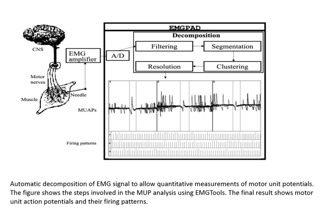
Resurgence of quantitative EMG was, however, aided by the development of computerized decomposition of the EMG signal which allowed extraction of MUPs which could then be measured by the computer. Several algorithms have been developed and are now installed on clinical EMG machines [6-8]. These advances allow rapid and accurate measurements of 20-60 MUPs from different sites in the muscle and have therefore made quantitation of EMG feasible in a busy clinical setting [9,10]. Quantitation of EMG may therefore become an integrated method in the differential diagnosis of muscle weakness. Furthermore, computerized quantitation of the interference pattern has been further developed to increase the diagnostic yield in patients with myopathy and neurogenic disease [11].
References
1. Adrian ED, Bronk DW: The discharge of impulses in motor nerve fibres: Part II. The frequency of discharge in reflex and voluntary contractions. J Physiol 1929, 67:i3-151.
2. Buchthal F, Rosenfalck P: Electrophysiological aspects of myopathy with particular reference to progressive muscular dystrophy. In Muscular dystrophy in man and animals. Edited by Bourne GH, Golarz MN: S. Karger; 1963:193-262.
3. Buchthal F: An introduction to electromyography. København, Stockholm, Oslo: Scandinavian University Books; 1957.
4. Rosenfalck P, Rosenfalck A: Electromyography – sensory and motor conduction. Findings in normal subjects. Copenhagen: Laboratory of Clinical Neurophysiology, Rigshospitalet; 1975.
5. Buchthal F, Kamieniecka Z: The diagnostic yield of quantified electromyography and quantified muscle biopsy in neuromuscular disorders. Muscle & Nerve 1982, 5:265-280.
6. Bischoff C, Stålberg E, Falck B, Eeg-Olofsson KE: Reference values of motor unit action potentials obtained with multi-MUAP analysis. Muscle Nerve 1994, 17:842-851.
7. Stålberg E, Falck B, Sonoo M, Stålberg S, Åström M: Multi-MUP EMG analysis – a two year experience in daily clinical work. Electroencephalogr.Clin.Neurophysiol. 1995, 97:145-154.
8. Nikolic M, Krarup C: EMGTools, an adaptive and versatile tool for detailed EMG analysis. IEEE Trans.Biomed.Eng 2011, 58:2707-2718.
9. Krarup C: Lower motor neuron involvement examined by quantitative electromyography in amyotrophic lateral sclerosis. Clin.Neurophysiol. 2011, 122:414-422.
10. Krarup C, Boeckstyns M, Ibsen A, Moldovan M, Archibald S: Remodeling of motor units after nerve regeneration studied by quantitative electromyography. Clin Neurophysiol 2015.
11. Fuglsang-Frederiksen A: The utility of interference pattern analysis. Muscle Nerve 2000, 23:18-36.
Dr. Christian Krarup works at the Department of Clinical Neurophysiology, Rigshospitalet and University of Copenhagen, Denmark


