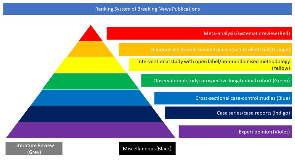
Cross-sectional case-control studies (Blue)
The authors conducted a longitudinal case-control 18F-FDG-PET study in patients diagnosed with COVID-19-related encephalopathy. Seven patients were followed-up at 3 timepoints: the first 18F-FDG-PET was performed in the acute phase, and consecutively at 1 month and 6 months. 18F-FDG-PET images were analyzed voxel-wise in comparison with 32 healthy controls. Patients’ neurological manifestations during acute encephalopathy were heterogeneous, but prominent cognitive and behavioral frontal disorders were ubiquitous. SARS-CoV-2 RT-PCR in the CSF was negative for all patients and MRI was unremarkable for most subjects. All patients had a consistent pattern of hypometabolism in a widespread cerebral network including the frontal cortex, anterior cingulate, insula and caudate nucleus, and mild hypermetabolism in the vermis, dentate nucleus and pons. One month later, hypometabolism was limited to the mediofrontal, right dorsolateral areas, olfactory/rectus gyrus, bilateral insula, right caudate nucleus and cerebellum, and no hypermetabolism was found. After 6 months, the majority of patients clinically improved but cognitive and emotional disorders remained, with attention/executive disabilities and anxio-depressive symptoms, coupled with lasting prefrontal, insular and subcortical 18F-FDG-PET/CT hypometabolism, even if less extensive. These findings could potentially explain the clinical features observed in patients with COVID-19, such as acute frontal lobe syndrome and subacute attention/executive deficits.


