presented by Vanessa Levrat & Anastasia Zekeridou
We report a case of a 76-year-old woman with a recurrent painful left ophthalmoplegia of obscure origin, with two episodes in two years.
The patient complains of pain in the left orbital area that worsens when she moves her eyes. She has diplopia which is maximal on looking to the left. There is no diurnal fluctuation. There are no other symptoms.
She presented with similar symptoms two years previously. The cerebral MRI, left temporal artery biopsy and blood workup at the time were unremarkable apart from a hypercholesterolemia. The diagnosis at the time was of a microvascular sixth nerve palsy in a patient with high blood pressure and hypercholesterolemia. She recovered completely after two months.
Presently, on examination, she showed an incomplete ptosis on the left with mydriasis (1mm of difference) and incomplete abduction of her left eye with horizontal diplopia which is maximal on abduction of the left eye without other signs. There was no proptosis, or scleral injection and no orbital bruit.
A neuroanatomical diagnosis of a partial third and sixth nerve palsies on the left was made.
The patient reported no significant family history.
The diagnostic work-up was comprehensive:
- There was no diabetes, no thyroid dysfunction or antibodies.
- The CK levels were normal and hypercholesterolemia was adequately controlled with oral treatment.
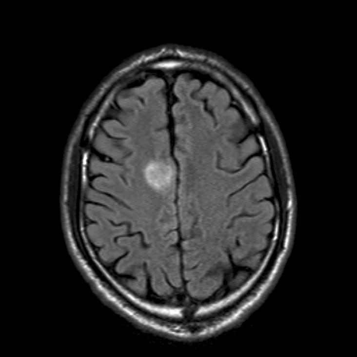
- The brain and orbital MRI and a thoraco- abdominal CT were normal.
- Clinical neurophysiological tests (ENMG) and anti-acetylcholine receptor antibodies were normal.
- The infectious and inflammatory blood screen (HIV, syphilis, tuberculosis, Lyme) were normal.
- A lumbar puncture showed a protein of 560 mg/ml.
After excluding an infectious disorder, the patient received oral prednisone treatment, which relieved her pain only.
Our final diagnosis was that of a painful ophthalmoplegia of unknown origin.
Dr. Vanessa Levrat and Dr. Anastasia Zekeridou both work at the Department of clinical neuroscience, Neurology Service, chaired by Prof. R. Frackowiak, CHUV Lausanne, Switzerland
Comment by the authors
We think that in our patient a recurrent neuropathy of microvascular origin is probable, given the hypercholesterolemia and treated hypertension, and absence of other abnormalities.
On the other hand, an inflammatory disease such as a Tolosa Hunt syndrome cannot be excluded given that 7.9% of these patients have no MRI findings (1).
Both of these disorders can be recurrent.
We do not believe that the patient has a myopathy or a neuromuscular junction disorder (pain, normal ENMG study and CK levels, normal MRI) nor an ophthalmoplegic migraine given the age and the absence of antecedent headache (2).
As for the long-term management, we have decided to gradually decrease prednisone doses.
A recurrence of symptoms would be in favour of Tolosa Hunt syndrome…REFERENCES:
1. S Colnaghi et al. ICHD-II Diagnostic Criteria for Tolosa-Hunt Syndrome in Idiopathic Inflammatory Syndromes of the Orbit and/or the Cavernous Sinus. Cephalalgia 2008 28: 577
2. L. La Mantia. Painful ophthalmoplegia: an unresolved clinical problem. Neurol Sci 2005 26:S79–S82
Comment by Silvia Colnaghi, Anna Pichiecchio, Maurizio Versino and Stefano Bastianello
This case report, by describing an interesting case of recurrent painful ophthamoplegia, demonstrates the difficulties embedded in the diagnosis of Tolosa Hunt Syndrome (THS) when the ICHD-II diagnostic criteria [1] (Table 1) are not fully satisfied.
In fact, the current THS diagnostic criteria are ambiguous regarding (i) the role of MRI (point B), and (ii) the dosage, schedule and expected effect of the steroid treatment (point D).
ICHD-II criteria are not mandatory about MRI methodology, but it is hard to reconcile the idea of THS as a possibly autoimmune, granulomatous disease with the possibility of a negative MRI as in this case report. Thereby, particularly in these cases, the MRI techniques should strictly adhere to the one proposed for evaluating the presence of granulomatous tissue in the cavernous sinus and/or the orbit, and a careful follow up should be mandatory both to confirm the THS diagnosis and to manage the steroid treatment [2]. More specifically, MRI should be performed with 3 mm thickness coronal and axial T1-SE and T2-TSE sequences and with T1 fat-suppressed weighted images after gadolinium administration at the level of the orbit and cavernous sinus.
Moreover, at least in cases with a negative MRI, the diagnostic work-up should also test, in addition to the full panel proposed by the authors, anti Gq1b antibodies to exclude a Miller-Fisher / Bickerstaff’ encephalitis [3].
Overall, since we do not have a biological marker for microvascular neuropathy (see point E), and the patient met the current THS diagnostic criteria, the diagnosis of THS could not be excluded, despite a recurrent microvascular cranial neuropathy was the most likely hypothesis.
1. Headache Classification Committee of the International Headache Society. Classification and diagnostic criteria for headache disorders, cranial neuralgias and facial pain.Cephalalgia 1988; 8 (Suppl. 7):1–96.1:
2. Colnaghi S, Versino M, Marchioni E, Pichiecchio A, Bastianello S, Cosi V, Nappi G. ICHD-II diagnostic criteria for Tolosa-Hunt syndrome in idiopathic inflammatory syndromes of the orbit and/or the cavernous sinus. Cephalalgia. 2008 Jun;28(6):577-84. Epub 2008 Mar 31. Review. PubMed PMID: 18384413.
3. Chiba A, Kusunoki S, Obata H, Machinami R, Kanazawa I. Serum anti-GQ1b IgG antibody is associated with ophthalmoplegia in Miller Fisher syndrome and Guillain-Barré syndrome: clinical and immunohistochemical studies. Neurology. 1993 Oct;43(10):1911-7. PubMed PMID: 8413947.Table 1: ICHD-II diagnostic criteria for the Tolosa-Hunt syndrome
13.16 Tolosa–Hunt syndrome
Description:
Episodic orbital pain associated with paralysis of one or more of the third, fourth and/or sixth cranial nerves which
usually resolves spontaneously but tends to relapse and
remit.
Diagnostic criteria:
A. One or more episodes of unilateral orbital pain persisting for weeks if untreated
B. Paresis of one or more of the third, fourth and/or sixth cranial nerves and/or demonstration of granulomas by MRI or biopsy
C. Paresis coincides with the onset of pain or follows it within 2 weeks
D. Pain and paresis resolve within 72 h when treated adequately with corticosteroids
E. Other causes have been excluded by appropriate investigationsDr. Silvia Colnaghi, Dr. Anna Pichiecchio, Dr. Maurizio Versino and Dr. Stefano Bastianello work at the C.Mondino National Institute of Neurology Foundation, IRCCS Pavia at the University of Pavia, Italy.
Comment by Michele Viana and Peter Goadsby
The authors present an interesting case of recurrent painful ophthalmoplegia. We agree with the authors that there are few similar cases in the literature described as “mysterious” –meaning a clear nosological entity accounting for the symptomatology was not identified.
Some considerations and comments:
On the presentation of the case we note that it was not reported whether the patient had an MRA or other angiography. The presence of a bruit is an important sign suggesting potential vascular causes, especially during the first approach to the patient, while its absence is not conclusive. Moreover, investigations for autoimmune presentations, such as SLE, sarcoidosis or Wegener’s Syndrome, would seem useful. It would have been useful to know if Gadolinium was given for the MRI. This can be useful to verify the presence of an inflammatory cranial neuropathy or a Tolosa-Hunt Syndrome. It might have been useful if some aspects of the pain were clarified, notably i) the quality and intensity of the pain; ii) the time relation between the onset of the ophthalmoplegia and pain; iii) the dosage and the schedule of prednisone therapy; iv) the time and extend of relief after the prednisone.
On the diagnostic discussion, a recurrent inflammatory cranial neuropathy seems a good possibility, given that condition often remits and relapses. An MRI with Gadolinium should be performed and more complete data about the effect of the corticosteroid therapy should be provided to verify this hypothesis. We would like to highlight that inflammatory cranial neuropathy is also now considered as the basis of ophthalmoplegic migraine, besides being associated to other conditions. “Ophthalmoplegic migraine” in fact it is a misnomer in that it is probably not a variant of migraine, at least in Caucasians, but rather a recurrent cranial neuralgia – in the second edition of the International Classification of Headache Disorders Ophthalmoplegic migraine was in fact classified under “cranial neuralgias” group. It is not correct to state that if the patient had no headache the diagnosis of ophthalmoplegic migraine is untenable.
A Tolosa-Hunt Syndrome (THS), which could fit with the clinical picture, has to be considered as a secondary option as an MRI should detect the typical changes. Yet, also here, we would like to know if the MRI was performed adequately to detect THS changes (with 3 mm thickness coronal and axial T1—SE and T2-TSE sequences and with T1 fat-suppressed weighted images after Gadolinium administration at the level of the orbit and cavernous sinus). In the case that a properly performed MRI was negative, the possibility that this patient is affected by a THS a very low. Just a very small part of patients with THS reported in literature (7.9%) had negative brain MRI. Moreover we should consider that those data came from cases studied with lower-field MRI with respect to what we use nowadays. Than this few patients (3 out of 64 described in the review cited by the Authors) might have presented structural changes that were not detected.
The microvascular hypothesis is less probable for the recurrence of the condition, in a patient with only hypercholesterolemia (well controlled) and high blood pressure as vascular risk factor, with no diabetes, no other signs of vascular lesions (either in the central nervous system or within other organs). In this context autoimmune serological test should be performed to exclude autoimmune diseases that might eventually cause microangiopathy. Anyway, also in such case, it would be difficult that such condition had involved just the microcirculation of two cranial nerves without giving other signs/symptoms or laboratory/imaging findings.
In conclusion we suggest making the patient have the exams not already performed. The therapy will come subsequently. With the data in our possession, assuming the patient is affected by a recurrent inflammatory cranial neuropathy, the steroid therapy is the more correct one. It should be noted that in such conditions although the pain usually respond to that therapy within few days, the neurological signs would probably take longer time to ameliorate.
Michele Viana is working at the Headache Science Centre, IRCCS “C.Mondino National Neurological Institute” Foundation, Pavia, Italy.
Peter Goadsby is Professor of Neurology at the Department of Neurology – Headache Group at the University of California, San Francisco, USA and President of the International Headache Society.


2 comments
Thanks for the very interseting case.Did you exclude Migraine? Sometimes migraine can present with ophthalmoplegia( painful type) without other classical features.
Regards
Shakya Bhattacharjee
Thank you for this interesting comment. We considered migraine but we excluded it given the age of onset (typically in younger patients) and the abscence of relative history of migraine in this patient.